EGAS AISovereign medical imaging · on the HOUSE engineEGAS AISovereign medical imaging · on the HOUSE engine
Egas is a sovereign, self-hosted medical-imaging mind built on the HOUSE AI engine. He reads a mammogram and outlines the lesion, reads a forearm X-ray and designs the printable cast that stabilises it, and tells a harmless mole from a possible melanoma — every read computed in his own Rust engine, on a plain CPU. No cloud, no external API, no data leaving your walls.
Named for Egas Moniz — the only Portuguese to win the Nobel Prize in Medicine — and, like him, built in Portugal.

Reads a 2D mammogram, localises the suspicious cluster and rebuilds it in an interactive 3D surgical view.
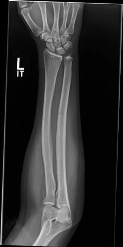
Open 3D view →Reads a forearm X-ray, flags the break, and designs a contoured 3D-printable orthosis and fixation plate.
See the fabricate flow →Snap or upload your own mole for an instant exemplar ABCDE read — in your browser. Demo only, not the real engine.
Try it on your mole →Egas interprets actual mammograms, telling benign tissue from malignant. Checked against public clinical data (CBIS-DDSM), the classifier he trained scores AUC 0.73 (0.77 on calcifications) — fair, honestly reported, and still climbing, not yet clinical-grade — and it runs entirely on your own hardware, on CPU, with no cloud and no GPU. The scans never leave the building.
Egas also outlines the lesion boundary, not just classifies it. Given the lesion’s location as a region prompt (the MedSAM protocol), he traces its outline at 0.847 mean Dice across 370 held-out discrete lesions — median 0.897, from patients never seen during training (biopsy-proven CBIS-DDSM, patient-disjoint split).
Masses 0.879 · calcifications 0.826 · malignant 0.854 vs benign 0.844 — no bias toward calling things benign. Accuracy falls off predictably on the smallest lesions rather than failing unpredictably.
The honest limit: this measures segmentation precision — how accurately Egas outlines a lesion he has been pointed at. It is not detection. Finding the lesion unaided, on a full mammogram, is the next stage and is not built yet. Research & capability demonstration — not a clinical diagnosis.
Finding the problem isn’t the end — Egas makes the tool that fixes it.
From a scan or a measurement, Egas designs a part in CAD, exports a printable mesh, and slices it into machine instructions — on his own hardware, no cloud CAD. Two real parts he made, for two different clinical problems:
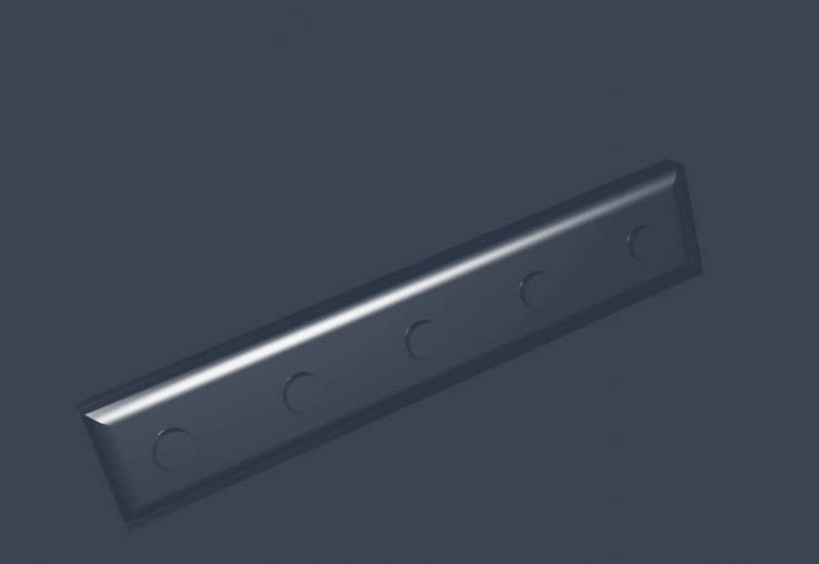
Osteosynthesis plate
A custom bone-fixation plate screwed to the bone. 62×12×3 mm, 5 countersunk holes → sliced to 25 layers, ~12 min, 715 mm filament.
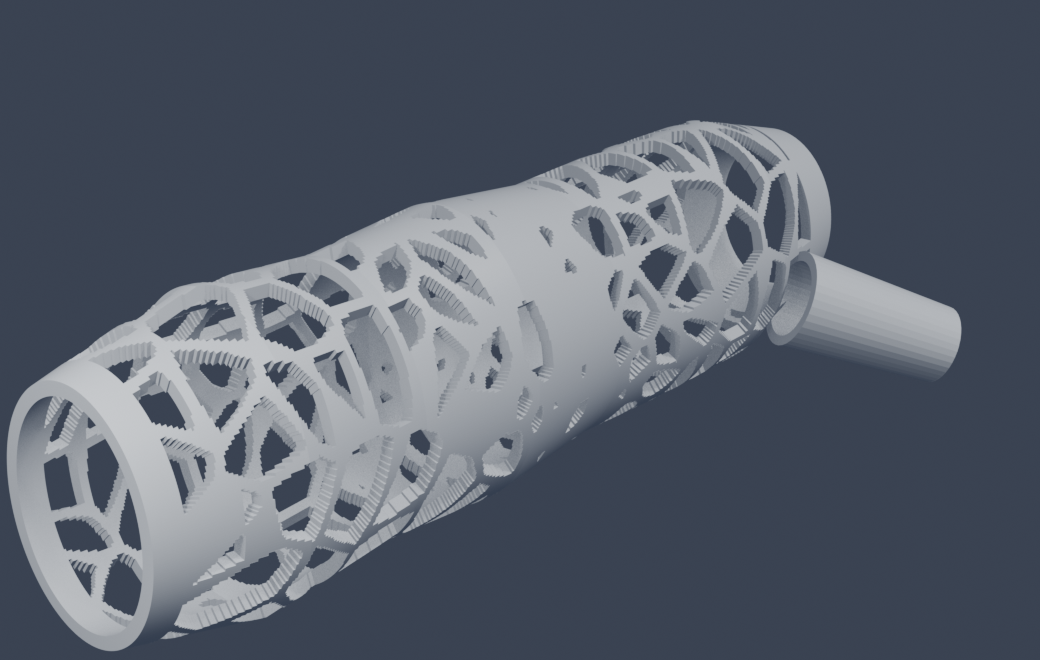
Contoured forearm–thumb cast
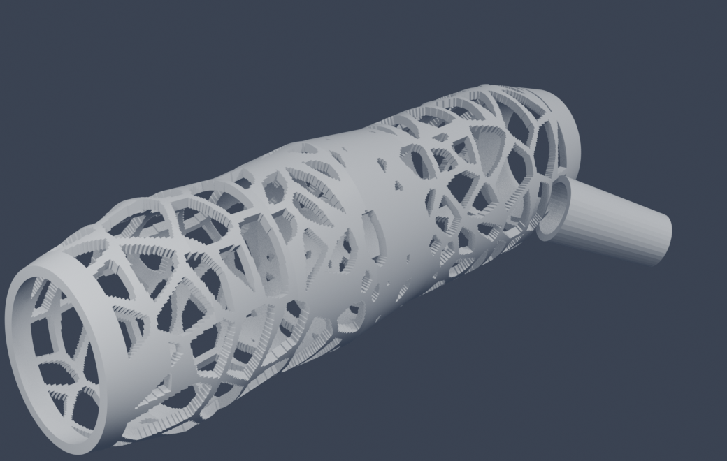
Reconstructed from the X-ray’s flesh silhouette at scale — a trabecular clamshell that follows the arm’s real contour, with a thumb sleeve that locks the wrist. Waterproof, breathable, no itch.
The cast follows the flesh — not a cylinder.
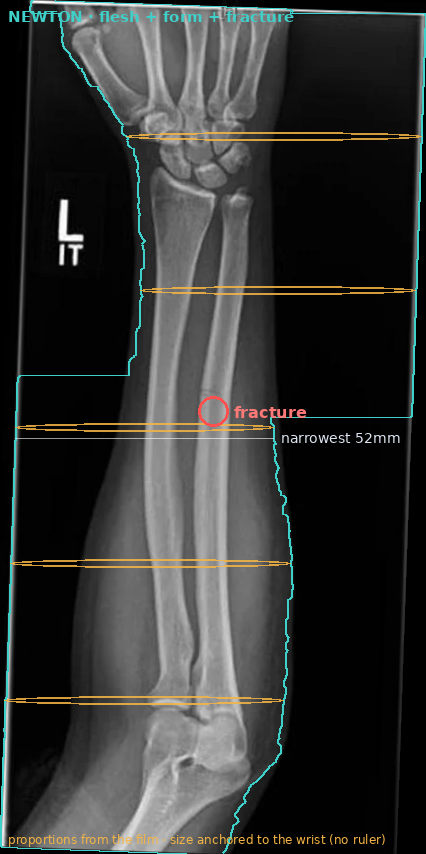
A plaster tube ignores the arm. Given a real forearm radiograph, Egas segments the soft-tissue silhouette, measures the width profile down the limb, flags the mid-shaft fracture by the bone-axis angulation, and rebuilds the forearm as elliptical cross-sections — then wraps it in a load-bearing trabecular mesh reinforced over the break. The shell is this arm’s own shape.
All three are real: the film is an actual both-bone forearm fracture; the middle tile is Egas’s own image processing on it (flesh segmented, form measured, fracture flagged); the cast is the mesh he generated from that read, ~340k triangles on-prem. Absolute size is anchored to the wrist (this film has no radiopaque ruler); fracture localisation is an early capability, not a clinical diagnosis.
And when the break needs to move: some fractures heal better with controlled wrist motion than with full lock-down — so Egas also builds a hinged variant, shown just below.
Research & capability demonstration — Egas reads the film and generates the parts on-prem; not certified medical devices, and fracture reading is an early capability, not a clinical diagnosis.
When the break needs to move: a cast with a wrist hinge.
Full immobilisation isn’t always best — some fractures recover faster if the wrist can still bend up and down. So Egas splits the cast at the wrist: a forearm sleeve and a hand piece, joined by a lateral hinge. The pivot lets the wrist flex; the boss-and-pin on each side blocks the rotation (the twist) that would destabilise the fracture. Controlled motion, not a rigid tube.
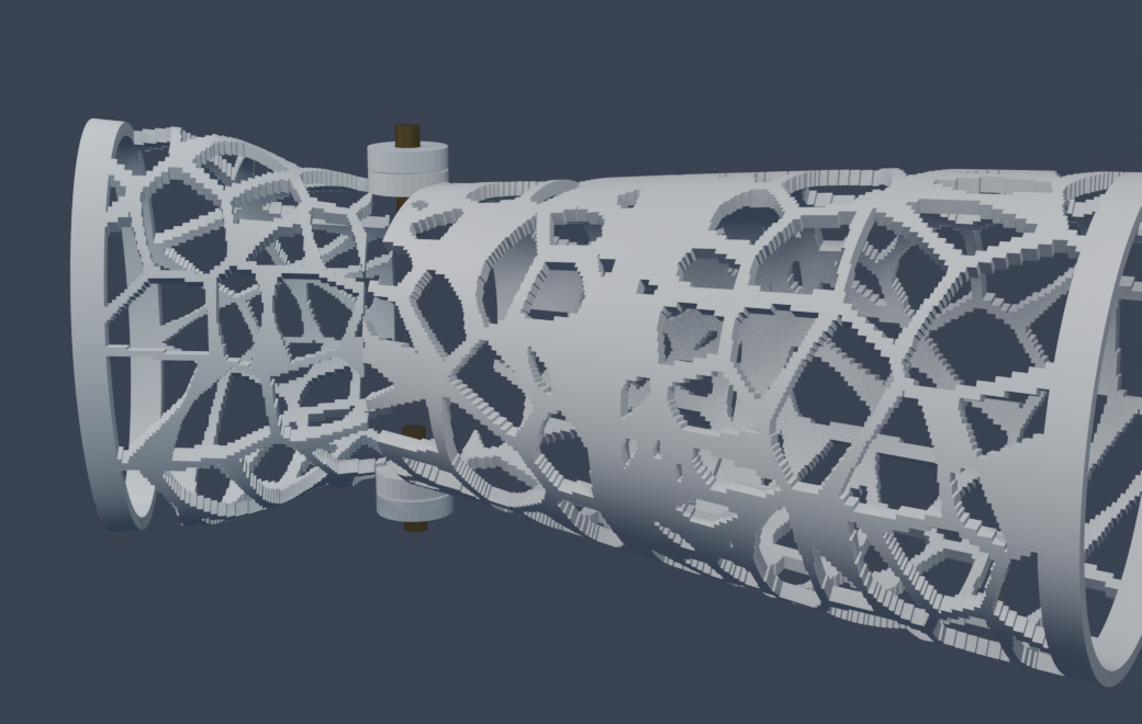
Shown mid-flexion — the hand piece (right) pivots on the wrist axle (amber pins) while the forearm sleeve (left) holds. Rotation about the arm’s long axis stays locked.
Research & capability demonstration — concept parts generated on-prem; not certified medical devices.
This morning Egas was set a new task: learn about skin moles and tell a harmless one from a possible cancer. It’s now mid-afternoon — and he already knows a couple of things about the craft. He can read a lesion, weigh it by its shape, border and colour, and flag a melanoma from an ordinary mole — running it on his own Rust engine, on-prem.
See what Egas learned about skin →Research & capability demonstration — not a certified medical device and not a clinical diagnosis. Every read is produced on-prem by the HOUSE engine; segmentation and fracture reading are early capabilities shown for evaluation, not for clinical use.